
Using Nanoparticles To Combat Arteriosclerosis
In industrialized countries, a high number of people suffer from arteriosclerosis – with fatal consequences: Deposits in the arteries lead to strokes and heart attacks. Researchers under the leadership of the University of Bonn have developed a method for guiding replacement cells to diseased vascular segments using nanoparticles. They demonstrated in mice that the fresh cells actually exert their curative effect in these segments. However, much research remains to be done prior to use in humans.
In arterial calcification (arteriosclerosis), pathological deposits form in the arteries and this leads to vascular stenosis. Strokes and heart attacks are a frequent outcome due to the resultant insufficient blood flow. Endothelial cells which line the blood vessels play an important role here.
“They produce nitric oxide and also regulate the expansion of the vessels and the blood pressure,” said junior professor Dr. Daniela Wenzel from the Institute of Physiology I of the University of Bonn. Damage to the endothelial cells is generally the insidious onset of arteriosclerosis.
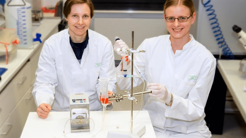
A team of researchers working with Junior Prof. Wenzel, together with the Technische Universität München, the Institute of Pharmacology and Toxicology at the University of Bonn Hospital and the Physikalisch-Technische Bundesanstalt Berlin, developed a method with which damaged endothelial cells can regenerate and which they successfully tested in mice. The scientists transferred the gene for the enzyme eNOS into cultured cells with the aid of viruses. This enzyme stimulates nitic oxide production in the endothelium like a turboloader.
“The enzyme is an essential precondition for the full restoration of the original function of the endothelial cells,” said Dr. Sarah Vosen from Prof. Wenzel’s team.
A magnet delivers the nanoparticles to the desired site
Together with the gene, the scientists also introduced tiny nanoparticles, measuring a few hundred nanometers (one-millionth of a millimeter), with an iron core.
“The iron changes the properties of the endothelial cells: They become magnetic,” said Dr. Sarah Rieck from the Institute of Physiology I of the University of Bonn. The nanoparticles ensure that the endothelial cells equipped with the “turbo” gene can be delivered to the desired site in the blood vessel using a magnet where they exert their curative effect. Researchers at the Technische Universität München have developed a special ring-shaped magnet configuration for this which ensures that the replacement cells equipped with nanoparticles line the blood vessel evenly.
The researchers tested this combination method in mice whose carotid artery endothelial cells were injured. They injected the replacement cells into the artery and were able to position them at the correct site using the magnet. “After half an hour, the endothelial cells adhered so securely to the vascular wall that they could no longer be flushed away by the bloodstream,” said Prof. Wenzel. The scientists then removed the magnets and tested whether the fresh cells had fully regained their function. As desired, the new endothelial cells produced nitric oxide and thus expanded the vessel, as is usual in the case of healthy arteries. “The mouse woke up from the anesthesia and ate and drank normally,” reported the physiologist.
Transfer to humans requires additional research
Normally, doctors surgically remove vascular deposits from the carotid artery and in some cases place a vascular support (stent) to correct the bottleneck in the crucial blood supply.
“However, these areas frequently become blocked with deposits once again,” said Prof. Wenzel. “In contrast, we are getting to the root of the problem and are restoring the original condition of healthy endothelial cells.” The researchers hope that what works in mice is also possible in humans, in principle.
However, there are still many challenges to overcome, Prof. Wenzel said, “There is still a considerable need for research.”