
Blind Woman Able To See Simple Shapes Using Brain Implant
Newly published research details how a team of scientists from the John A. Moran Eye Center at the University of Utah and Spain’s Miguel Hernández University successfully created a form of artificial vision for a blind woman using a prosthesis hardwired into her brain.
Publishing “Visual percepts evoked with an Intracortical 96-channel Microelectrode Array inserted in human occipital cortex” in The Journal of Clinical Investigation, Moran researcher Richard A. Normann, PhD, and Spanish collaborator Eduardo Fernández, MD, PhD, detail how the Moran|Cortivis Prosthesis produced a simple form of vision for 60-year old Berna Gómez. The team conducted a series of experiments with Gómez for six months in Elche, Spain, that represent a leap forward for scientists hoping to create a visual prosthesis that could increase independence for the blind.
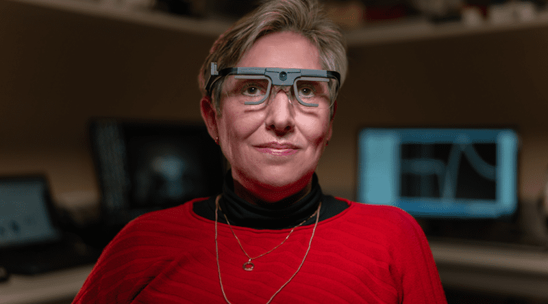
A neurosurgeon implanted a microelectrode array invented by Normann, the Utah Electrode Array (UEA), into the visual cortex of Gómez to both record and stimulate the electrical activities of neurons. Gómez wore eyeglasses equipped with a miniature video camera; specialized software encoded the visual data collected by the camera and sent it to the UEA. The array then stimulated neurons to produce phosphenes, perceived by Gómez as white points of light, to create an image.
A former science teacher fully blind for 16 years at the time of the experiments, Gómez had no complications from the surgery and researchers determined that the UEA did not impair the function of neurons in close proximity to the electrodes or affect the function of the underlying cortex. Gómez was able to identify lines, shapes, and simple letters evoked by different patterns of stimulation. To help her practice using the prosthesis, researchers created a video game for Gómez to play using a character from the popular television show The Simpsons. Thanks to her precise descriptions of visual perceptions and importance to the research, Gómez is a co-author on the study.
“These results are very exciting because they demonstrate both safety and efficacy,” said Fernández, who has collaborated with Normann for more than 30 years and is an adjunct professor at Moran. “We have taken a significant step forward, showing the potential of these types of devices to restore functional vision for people who have lost their vision.”
Normann and colleague Gregory Clark, PhD, first used the UEA in amputees to evaluate its safety and efficacy. The UEA allowed the amputees to control artificial limbs simply with their desire to move a finger or hand. The Spain experiments were the first to implant the UEA into the visual cortex.
The team’s neurosurgeon placed just one 4 mm by 4 mm UEA in Gómez’s brain, but published research conducted by Normann indicates between seven and 10 arrays in the visual cortex, working together, could produce more detailed images for useful vision. University of Utah neurosurgeons and neuroscientists John D. Rolston, MD, PhD, and Tyler Davis, MD, PhD, collaborated on the research.
“One goal of this research is to give a blind person more mobility,” said Normann. “It could allow them to identify a person, doorways, or cars easily. It could increase independence and safety. That’s what we’re working toward.”
The research team hopes the next set of experiments will use a more sophisticated image encoder system, capable of stimulating more electrodes simultaneously to reproduce more complex visual images.